SimedoBrowse
Sign inUpload
Simedo
AboutDocsGuidelinesPrivacyTermsContactGitHub

© 2026 Simedo · See molecules in motion

Browse simulations

Filter by category, family, organism, and more.

Showing 11 of 20 simulations

Filters

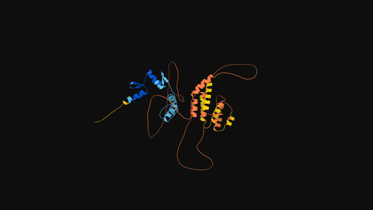
ZSCAN4, zinc finger and SCAN domain-containing protein 4

Predicted full-length structure of human ZSCAN4 (433 residues), a zinc finger transcription factor with a SCAN dimerization domain and tandem C2H2 zinc finger array. ZSCAN4 is transiently expressed in two-cell-stage embryos and in a small subpopulation of mouse embryonic stem cells, where it has been implicated in telomere maintenance and genome stability during the totipotent state.
ProteinZinc finger proteins
Source: RCSB PDB

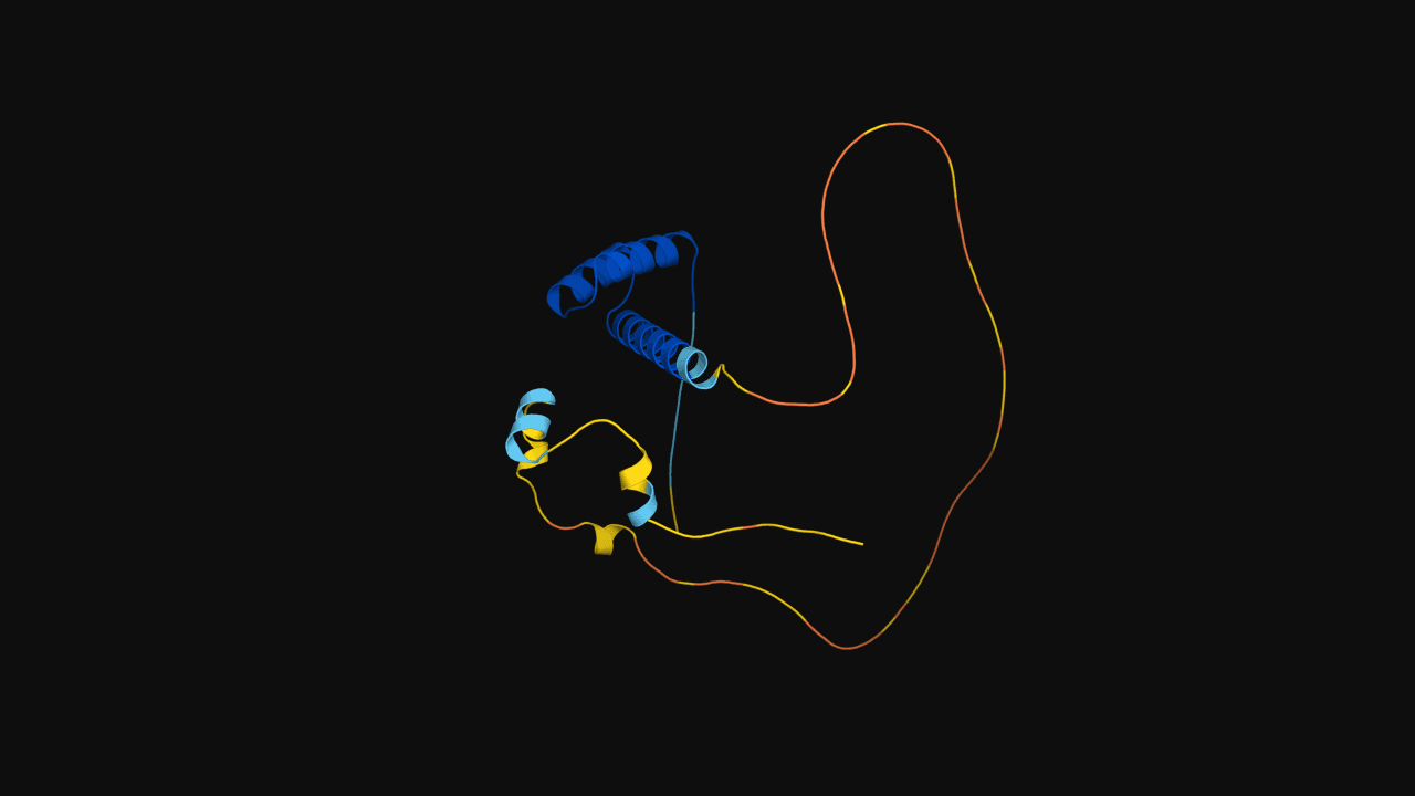
LEUTX, paired-like homeodomain transcription factor

Predicted full-length structure of human LEUTX (198 residues), a paired-like homeodomain transcription factor expressed transiently during the first cleavage divisions of the human embryo. LEUTX is part of the embryonic genome activation network and its expression is rapidly silenced after the eight-cell stage.

ProteinHomeodomains
Source: RCSB PDB

DUX4 double homeodomains bound to target DNA

Crystal structure of the human DUX4 transcription factor's tandem homeodomains in complex with a DNA duplex containing the consensus binding motif. Homeodomain 1 recognizes the TAA element via the canonical helix-in-major-groove contact, while homeodomain 2 reads the TGA element. DUX4 is the master regulator of zygotic genome activation in human embryos and is causally implicated in facioscapulohumeral muscular dystrophy (FSHD).

ProteinHomeodomains5ZFZ
Source: RCSB PDB

Oxyhemoglobin, R state

Human hemoglobin in the oxygen-bound R state. The classic structure that captures cooperative binding mid-cycle.

ProteinGlobins1HHO
Source: RCSB PDB

SARS-CoV-2 spike, closed prefusion

Closed-trimer prefusion conformation of the spike glycoprotein. The starting state for receptor-binding-domain opening dynamics.

ProteinCoronavirus spike6VXX
Source: RCSB PDB

Sperm whale myoglobin

The first protein structure ever solved by X-ray crystallography (Kendrew, 1958). Still a benchmark for oxygen-storage dynamics.

ProteinGlobins1MBN
Source: RCSB PDB

Deoxyhemoglobin, T state

Human deoxyhemoglobin in the tense T state. Pairs naturally with 1HHO to study the allosteric switch behind cooperative oxygen binding.

ProteinGlobins4HHB
Source: RCSB PDB

Crambin

A 46-residue plant protein resolved at sub-ångström resolution. A tiny but exceptionally well-resolved benchmark for force fields.

Protein1CRN
Source: RCSB PDB

Nucleosome core particle

147 base pairs of DNA wrapped around a histone octamer (two copies of H2A, H2B, H3, H4). The fundamental unit of chromatin.

ProteinHistones1KX5
Source: RCSB PDB

Bovine pancreatic trypsin inhibitor (BPTI)

Small, exceptionally stable Kunitz-domain inhibitor of serine proteases — one of the most thoroughly studied proteins in biophysics.

ProteinKunitz inhibitors4PTI
Source: RCSB PDB

Ubiquitin

Seventy-six residues of fold-and-signaling perfection. The reference β-grasp fold and a staple of folding studies.

ProteinUbiquitin1UBQ
Source: RCSB PDB